


Nanoparticles in medicine
Medicine is pinning its hopes on nanoparticles. These are used in cancer treatment, as MRI contrast agents and in vaccine development. But what exactly do these tiny particles do inside the body? In a project funded by the German Research Foundation (DFG), researchers at h_da are exploring this very question by developing tiny model kidneys and dispatching nanoparticles of various shapes – sometimes round, sometimes star-shaped, sometimes spiky – through them. The aim is to gain a better understanding of how nanoparticles work in the body and to minimise side effects.
By Christina Janssen, 1.6.2026
Some stories are fascinating from start to finish – but unexpected in a chemistry lab: tiny gold stars under an electron microscope, millimetre-sized model kidneys on plastic chips, cells that devour anything and everything that causes disruption in the body, and substances that can dissolve metal, glass and bone. Yet all this is currently coming together in h_da’s research laboratories – in a nanoparticle project funded by the German Research Foundation (DFG).
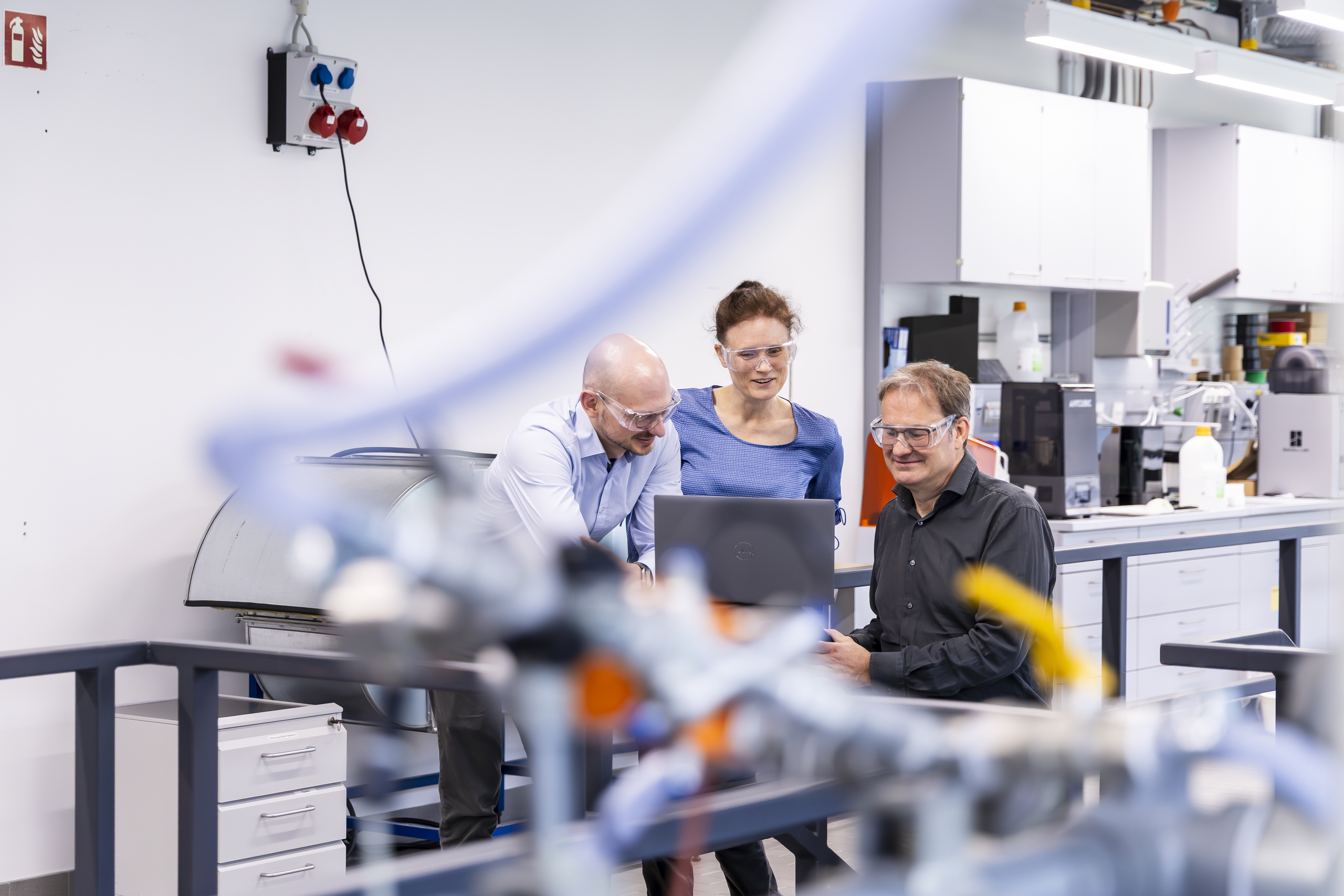
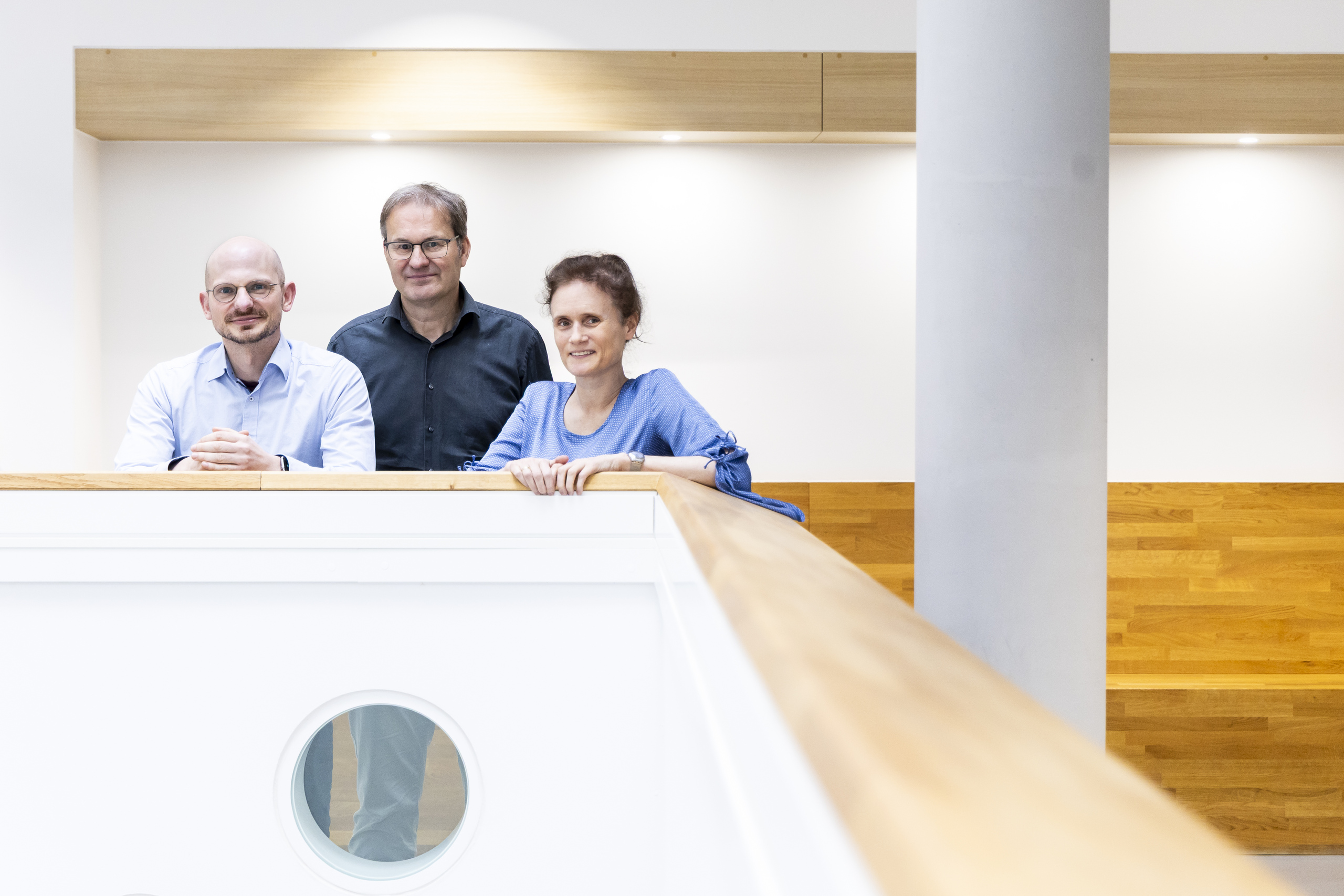
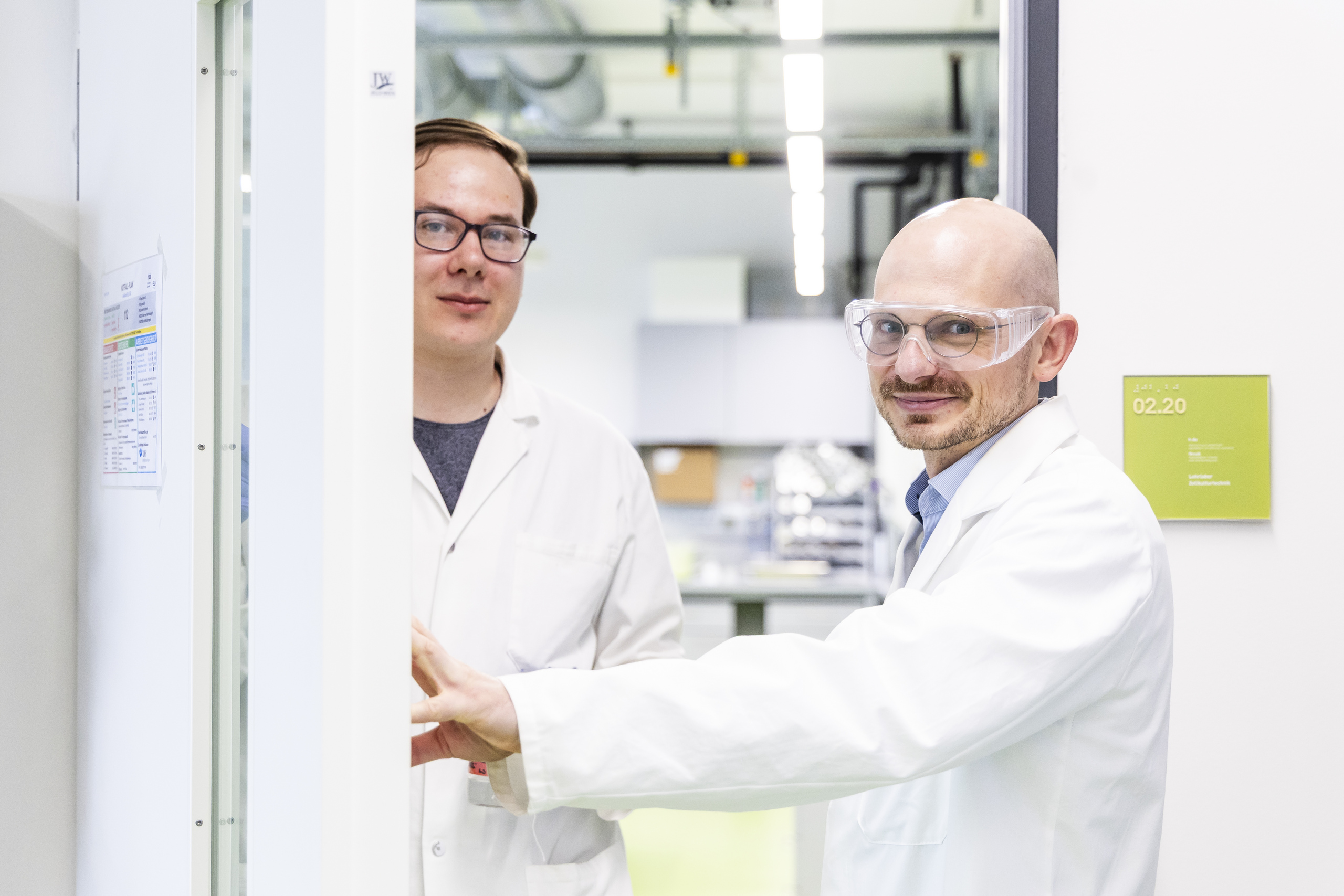
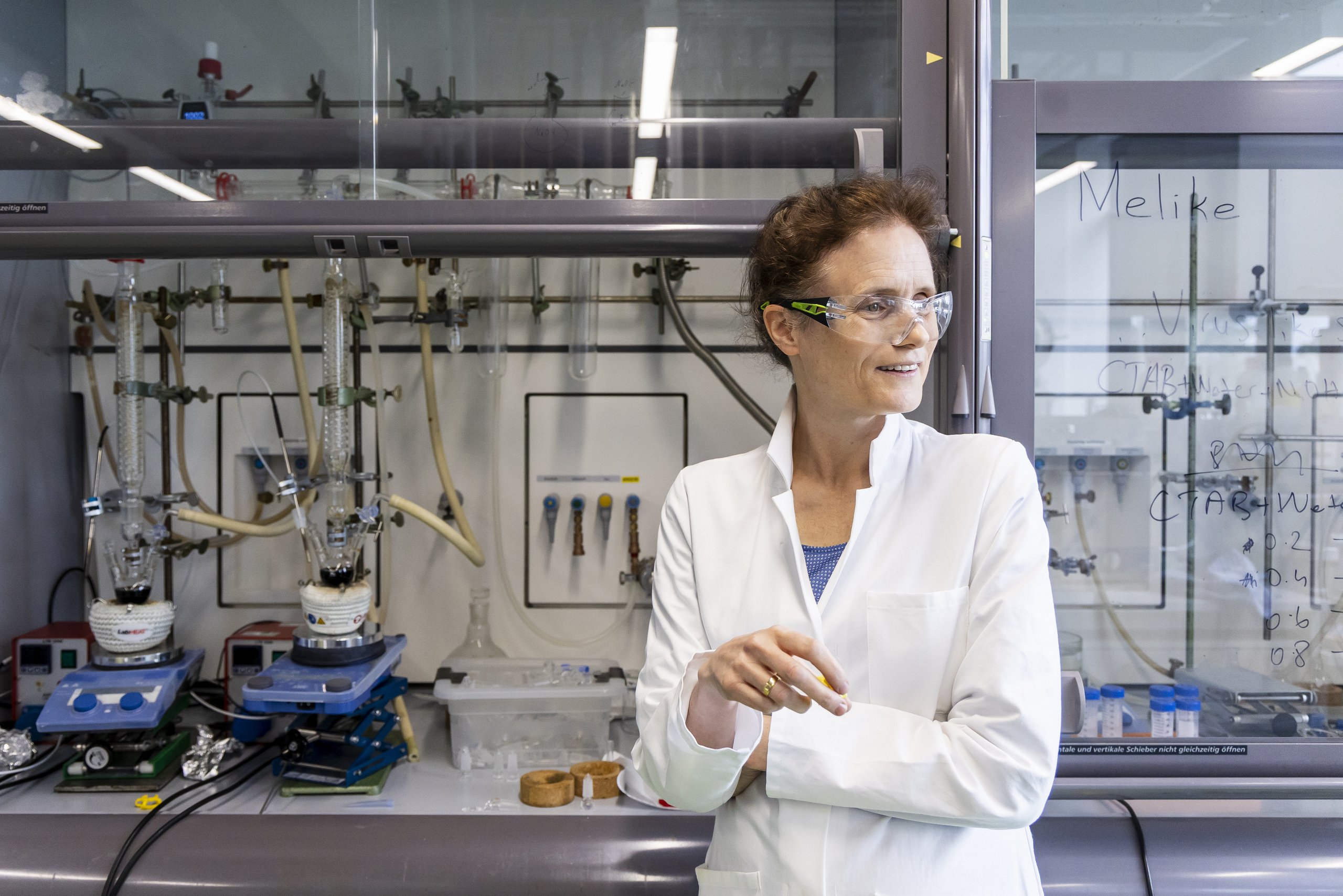
“Nanoparticles are between one and one hundred nanometres in size,” explains Professor Christina Graf from the Faculty of Chemical Engineering and Biotechnology. “A nanometre is 10⁻⁹ m, or one billionth of a metre.” Put simply: nanoparticles are extremely small. Some are luminescent, others are magnetic, while yet others are suitable vehicles for transporting drugs directly to tumour cells. That is why research is currently devoting so much attention to these tiny particles. Alongside nanoscientist Professor Christina Graf, the h_da team includes Professor Michael Becker, a cell biologist, and Professor Frank Schael, an expert in microprocess engineering.
Little helpers with great potential
Lots of people come into contact with nanoparticles without realising it – for example, during an MRI scan. Such scans use iron oxide nanoparticles as a contrast agent. Gold nanoparticles are also regarded as promising little helpers in medicine. “They are used for photothermal therapy in cancer treatment,” says Professor Graf. Here, the gold nanoparticles act as “thermal amplifiers” to destroy tumour cells through heat. Anyone assuming, however, that the faculty’s basement houses crates of gold bars is mistaken: “We will indeed also work with gold particles, but not with piles of them,” Graf makes clear.
But before newly developed particles can be used in practical applications, science needs to understand how they behave inside the body. This is where things get complicated: nanoparticles can clump together, accumulate, or end up in the wrong place. The result is side effects such as cell damage or death. This especially occurs in the kidneys. “The kidneys are exposed to a lot of toxic compounds,” explains cell biologist Professor Michael Becker. After all, their job is to filter out many substances that stress and disrupt the body – including the contrast agent that a patient must flush out by drinking large amounts of water after an MRI scan.
A kidney-on-a-chip
To gain a better understanding of the risks posed by nanoparticles, the h_da researchers are creating “tiny kidneys” from different types of cells and using them in their experiments as models that are as close to the physiology of a real kidney as possible. How do fluids and particles flow through the blood vessels? Where do nanoparticles become trapped? Where do clumps form? And exactly how does cell damage occur? The team is able to visualise all this under the high-resolution fluorescence microscope that h_da was also able to purchase with funding from the German Research Foundation. Professor Frank Schael names one of the key advantages of this set-up: “We can dispense with human subjects and animal testing in our experiments.”
The magic solution: organ-on-a-chip. “The tiny organs form on specially designed plastic chips,” explains Professor Frank Schael, who develops the chips himself and uses 3D printing to make them in the lab. They are actually small plastic boxes in which cells are cultivated to form three-dimensional structures – a big difference to research conducted with conventional, two-dimensional cell cultures: “The cells form tube-like structures – similar to blood vessels or renal tubules in the human body,” explains cell biologist Becker. To do this, the researchers skilfully combine three types of cells: kidney cells, which are particularly prone to damage, blood vessel cells and immune cells. “The third are phagocytes that devour everything that causes disruption in the body,” says Becker. That includes nanoparticles.
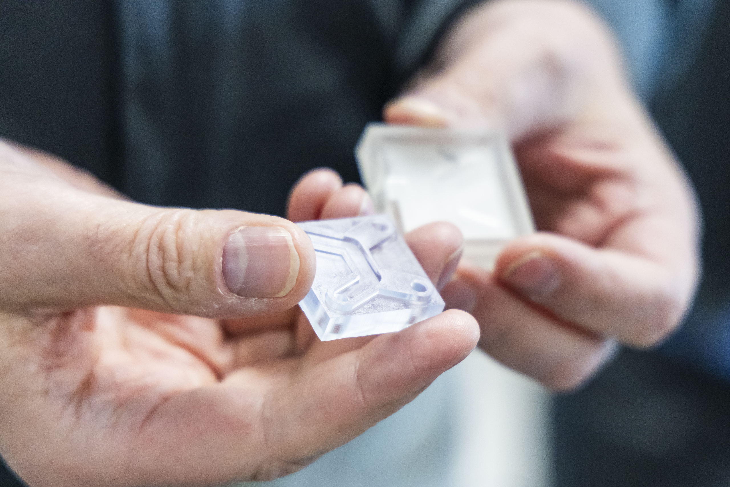
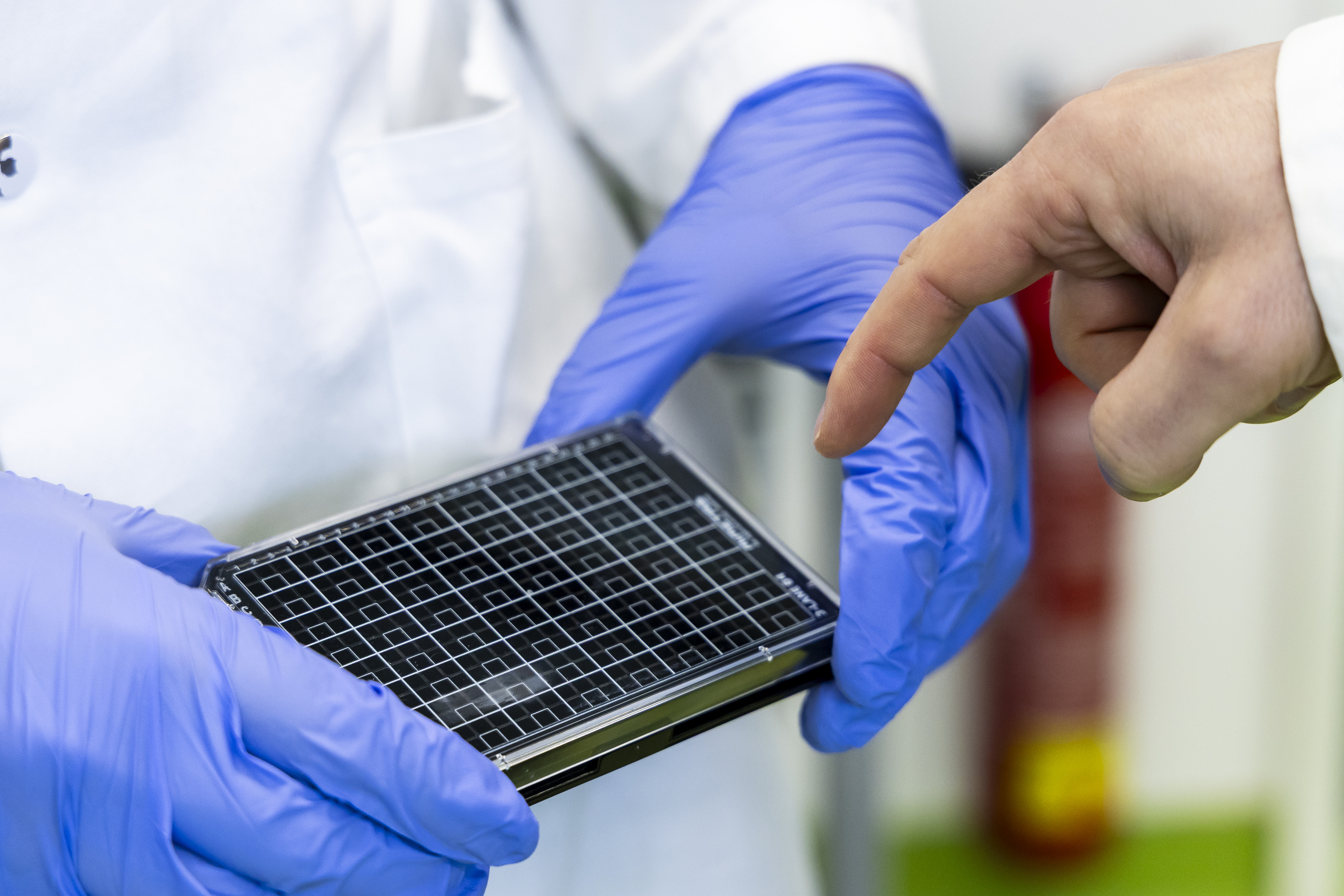

The chip is equipped with fine channels, through which fluids containing nanoparticles are directed inwards and flow through the kidney tissue – a highly complex process almost like in real life, as things never come to a halt in a real kidney either: blood flows through a network of channels, branches and bends. “Why has nature designed the kidney exactly like this, and what does it mean for nanoparticles’ mode of action?” says Schael, describing one of the project’s main questions.
Dead cells under the microscope
The researchers additionally want to find out how the material, shape, size and surface structure of nanoparticles influence their toxicity. And how many nanoparticles actually reach their intended target. “Christina is passionate about nanoparticles,” jokes cell biologist Becker. “And I’m more passionate about cells.” That’s why the team is examining both: Do the nanoparticles reach their intended destination? And do more or fewer cells die off?
Michael Becker is able to analyse the effects very accurately in his laboratories: using various staining methods, in the first step he determines whether the kidney tissue is growing properly on the chips. After experiments with various nanoparticles, the extent of tissue damage becomes visible under the microscope: “The question is, quite simply: Do more or fewer cells die off if the shape of the nanoparticles changes in any way?” says Becker.
Spheres, stars, spikes – and Christmas card motifs
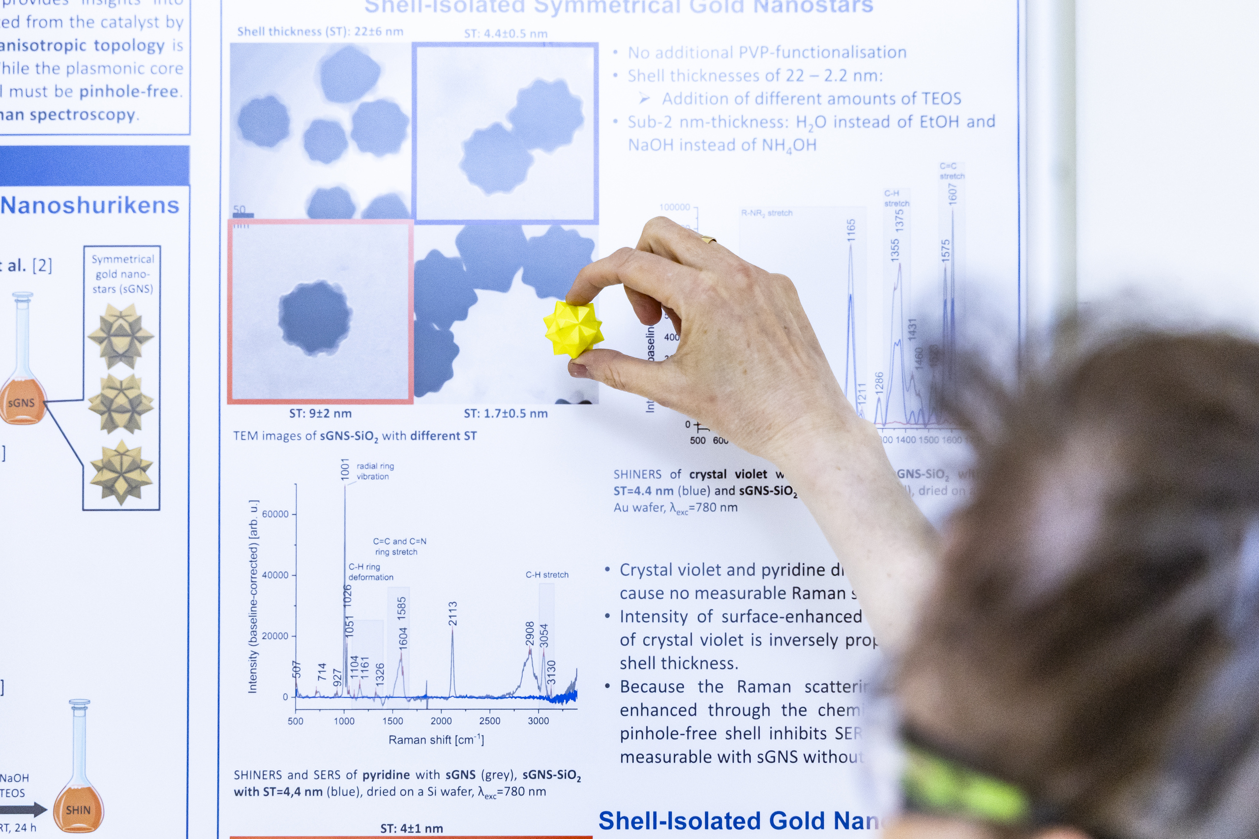
There is no end to Professor Graf’s inventiveness: in her laboratories, she produces the required nanoparticles in a wide variety of shapes. “We have spherical nanoparticles,” she says. “But we also have particularly long ones, as well as cube-shaped and triangular ones. Some resemble little stars.” The shape is not “just for fun”. It directly influences how the particles move around in the body. “If they simply roll, it’s easy,” explains Graf. Spiky particles, by contrast, become trapped more easily, or cells absorb them more quickly.
In the first instance, the production of the nanoparticles is a classic “cooking” process in the laboratory: “They are synthesised in small quantities in glass flasks,” says Graf drily. Although that sounds simple, the result is not random particles, but precisely designed nanostructures. “You can block individual surfaces so that the nanoparticles grow into a specific shape – for example, as rods or cubes.” The particles then look surprisingly ornate under the electron microscope.

Graf is particularly proud of the gold icosahedral stars created by Daniel Nachtsheim, her doctoral student. “They were even used for a Christmas card last year,” she is pleased to report. But there is also a scary side to her laboratory: to dispose of the nanoparticles after use, large containers of highly corrosive substances are stored in a fume cupboard there. They go by names such as “aqua regia” or “piranha solution”, can even dissolve gold, bone or glass – and sometimes (much to Graf’s amusement) interest visitors more than the extremely expensive equipment around them.

The overarching goal: better therapies
At the end of the day, the central question is: How can nanoparticles for medical applications be designed so that they are as useful and safe as possible? In Becker’s words: “If we could ultimately file a patent, that would be a dream result. If, in the end, we could know exactly how nanoparticles need to be shaped so that toxicity in the kidneys is reduced by 50%, for example.” This would make it possible in the future to use higher concentrations of contrast agents, making MRI scans more precise and thus more conclusive. But fundamental insights would be valuable too, such as why certain cells react differently to others under specific flow conditions.
Even if no new medical applications are on the immediate horizon, the project could contribute to a better understanding of the risks – in areas outside medicine as well. After all, it is not only in the lab that we come into contact with nanoparticles. “We inhale them, absorb them via our skin and ingest them with our food,” says Professor Graf. “They are found in exhaust fumes, dust and cosmetics.” In a nutshell: these tiny particles have long been everywhere, and now the h_da researchers are trying to find out when they are little helpers – and when they are troublemakers. A question evidently of interest to NASA too. Organ-on-a-chip systems were already used on the latest Artemis mission. Although the h_da researchers are not yet working on space travel, they are nevertheless helping to advance technologies with a far-reaching impact.
Related articles
impact, 20.04.2025: CANCER, PARKINSON'S DISEASE, AGEING PROCESSES
impact, 18.11.2024: Michael Becker ist Professor des Jahres 2024 (in German only)
impact, 18.06.2024: "A BOOST FOR JOINT RESEARCH"
impact, 29.01.2024: SUSTAINABLE DRUG DEVELOPMENT
impact, 10.01.2022: HUNTING DOWN PROTEINS
Contact our Editorial Team
Christina Janssen
Science Editor
University Communications
Tel.: +49.6151.533-60112
Email: christina.janssen@h-da.de
Translation: Sharon Oranski
Photography: Samira Schulz
Study programmes
MSc in Chemical Engineering and Biotechnology
BSc in Biotechnology (in German only)
BSc in Technical Chemistry (in German only)
Dual study programmes (in German only)