


New approach in the diagnosis of Alzheimer’s disease

At the present time, around 1.8 million people in Germany are living with dementia, most of whom have Alzheimer’s disease. According to forecasts, the numbers will rise significantly unless there is a medical breakthrough in the next few years. Still, the first drug that specifically targets the protein deposits in the brain that are characteristic of Alzheimer’s disease was approved in the USA last year. However, the new drug only helps if the disease is detected at an early stage. This is the starting point for a team from the Faculty of Mathematics and Natural Sciences at Darmstadt University of Applied Sciences: in a project funded by the EU, the h_da researchers are working with partners in the Netherlands, the UK and Germany on new methods for diagnosing Alzheimer’s disease. Their aim is to detect the disease earlier and treat it more successfully – as soon as suitable drugs come onto the market.
By Christina Janssen, 13.2.2024
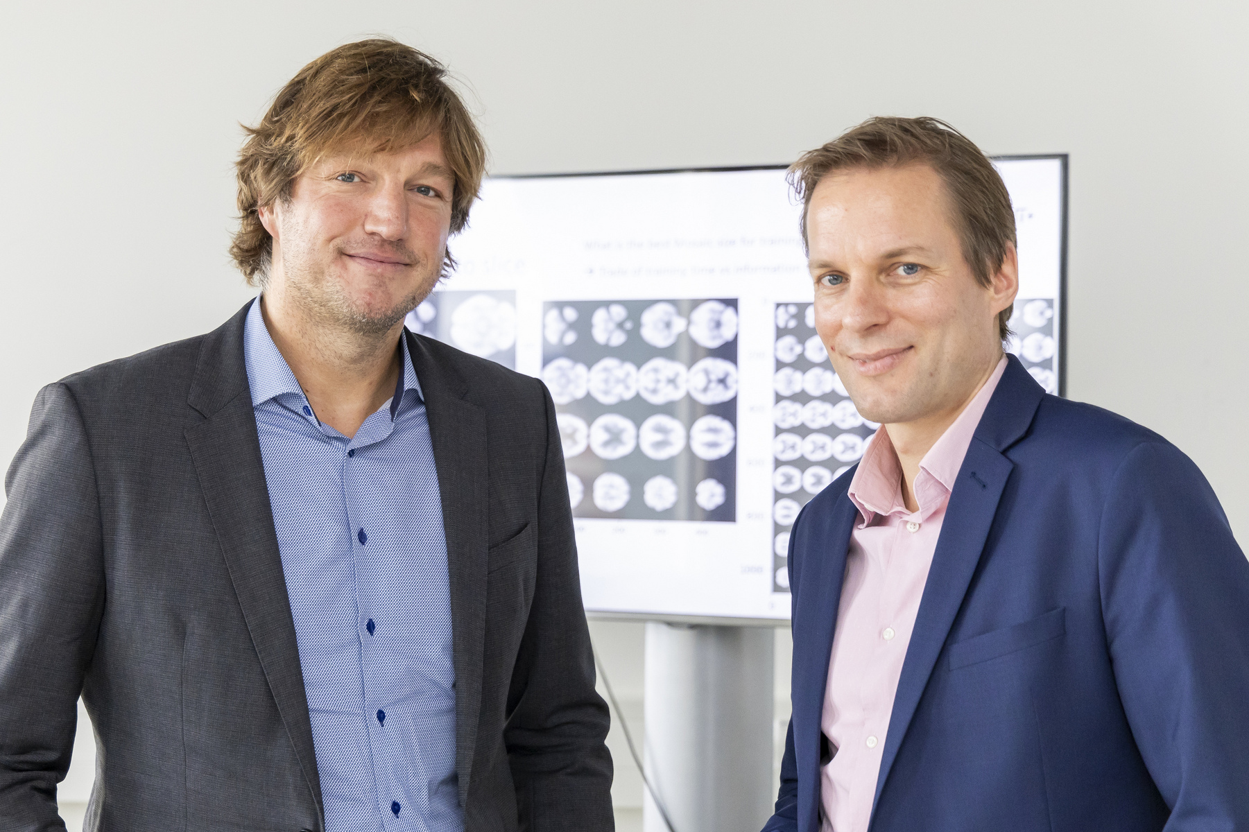
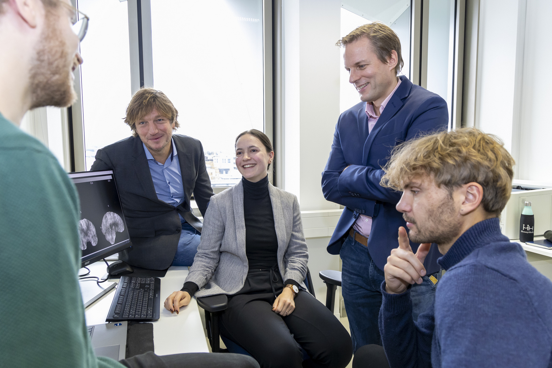
You could call the research project “Resist beginnings!”, but in fact it has a slightly bulkier title: “Arterial spin labelling Scanner- and Patient-Independent Robust diagnostic Evaluation” (ASPIRE for short). The project aims to improve the treatment prospects for Alzheimer’s patients in the long term, a hope nurtured by millions of people – both patients and their relatives. Physics professor Johannes Gregori and mathematics professor Andreas Weinmann have joined forces for the project. Their team also includes doctoral researcher Chiara Schmidt as well as Master’s students Ben Isselmann and Jakob Seeger. They are collaborating within the project, which is funded by the EU, with Amsterdam UMC (University Medical Centre), mediri, a company based in Heidelberg that specialises in medical imaging, and Gold Standards Phantoms, a British manufacturer of medical technology devices.
The researchers have tasked themselves with finding a new approach to the diagnosis of Alzheimer’s disease. Today, the disease is usually diagnosed through conventional MRI brain scans – in addition to cognitive assessments and laboratory tests to determine whether disease-specific proteins are present. This standard procedure maps the status quo of the brain. A reliable method, but not yet the best, according to the scientists in Darmstadt: “With this method, it is only possible to detect changes in the brain at a very late stage,” explains Professor Johannes Gregori. “If, by contrast, you look at changes in metabolism or blood flow in the brain at an earlier stage, there is a good chance of detecting the disease earlier.”
Medical examinations without contrast media
That is why the research team at h_da is looking very closely at two other diagnostic methods. One is positron emission tomography (PET). In the variant under study, it images changes in the brain’s blood glucose metabolism, which can be a very early sign of Alzheimer’s disease. This is an established method, but the examination is hard on the body because of the need to administer a slightly radioactive contrast medium. And it is costly – around USD 700 per scan. By contrast, the second method that Gregori, Weinmann & Co. have in mind has significant advantages: “Arterial Spin Labelling (ASL)”, which is also referred to in the project title, is many times cheaper, does not require a contrast medium and is equally able to show early indications of disease. ASL is a special variant of MRI, but it does not image a static condition. Instead, it shows changes in blood flow in the brain: How much blood flows in and out – and how quickly? These parameters can also be indicators of early-stage Alzheimer’s disease.
In combination with an AI system, this method so far hardly used could become the new “gold standard” in the diagnosis of Alzheimer’s disease. “Expensive and stressful examinations would not normally be carried out on healthy people or patients without symptoms,” says physicist Gregori. “ASL scans, by contrast, are far cheaper and could even become part of routine screening at a younger age.” As yet, there is no hope of a cure for Alzheimer’s disease, but drug research to remedy this situation is being conducted worldwide. Lecanemab (marketed under the name “Leqembi”), which was approved in the US in 2023 for treating Alzheimer’s disease, can slow disease progression in the early stages. The more drugs of this type that come onto the market, the more important it becomes to diagnose the disease as early as possible. “As soon as an effective therapy for Alzheimer’s disease becomes available, doctors must be able to detect the disease as quickly as possible so that treatment can then start. This is what we’re working on. Even if it’s still a distant prospect for the vast majority of people currently affected.”
AI-supported diagnosis
But how early is ‘as early as possible’? “Six years,” says Professor Gregori. “There can be indications even six years before the final diagnosis that Alzheimer’s disease is highly likely to develop.” Thanks to the AI-supported analysis of the PET images described above, this is already possible today. The h_da team is now striving for the same or even better results with the more health-friendly and cost-saving ASL scans. “It will be exciting to see if we are successful,” says Gregori.
Specifically, the h_da team would like ASL images to deliver diagnoses of Alzheimer’s disease that are just as reliable as the standard methods used today. This is a challenge for various reasons. Ben Isselmann and Jakob Seeger, who were responsible within the project for the acquisition and technical processing of vast amounts of image data, describe the baseline as follows: “It’s not so easy to obtain ASL images because the method is still relatively new. In addition, the images have to be extensively pre-processed before we can use them.” This is because fluctuations in ASL measurements are very high in comparison to other methods, meaning that images for the same patient – but taken at different times – can vary considerably: “Whether a patient drinks coffee or smokes before the scan, exercises or doesn’t, makes a difference to blood flow.” This makes it all the more difficult to extract the typical characteristics of Alzheimer’s disease from the data.
Uncharted scientific territory
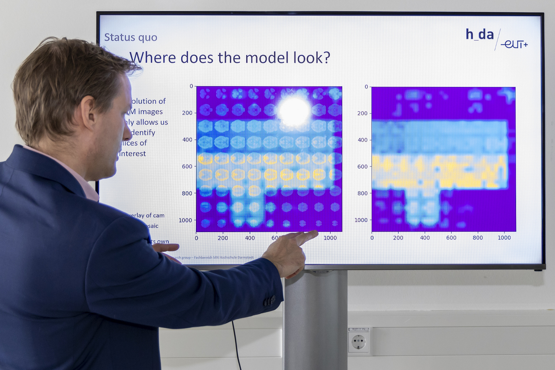
“This is where artificial intelligence comes into play,” explains doctoral researcher Chiara Schmidt. The underlying principle is that the AI recognises the patterns characteristic of Alzheimer’s disease in images and on that basis suggests a diagnosis – Alzheimer’s disease, not Alzheimer’s disease, or an intermediate form. The AI system developed by the h_da team and trained with vast numbers of datasets already performs this task very efficiently with MRI and PET data: a first success for the researchers in Darmstadt. “The next step is to transfer this to the ASL data,” reports Chiara Schmidt. “This is the point where we enter uncharted scientific territory – and where the biggest challenge lies.”
Uncharted scientific territory also means that the team is working with technology at the highest level. “We use what is known as a transformer model,” explains AI expert Andreas Weinmann. "This is the latest craze, so it really is a ‘hot-off-the-press model’. The results it delivers are comparable to – and partly even better than – the state of the art in medicine. I don’t see how this can be achieved with conventional methods.”
“Firework of methods”
The “transformer” used by the researchers analyses images using a method similar to that which ChatGPT uses to produce texts – or like the DALL-E programme, which generates images from spoken sentences. “The first revolution in this area was neural networks on the basis of ‘convolutional modules’. These are faster, but not as precise as transformers. Convolutional modules are based on approaches previously used in conventional image processing,” explains Andreas Weinmann. “With the new transformers based on ‘attention modules’, we’re now experiencing the second revolution: although these transformers need more computing power and are slower, they are more accurate.” Technological progress is happening at an accelerated pace, he adds. “Innovations are coming in quick succession. On the one hand, it’s fantastic that new methods are emerging in such a short space of time. On the other hand, it’s hard to keep up. It’s a firework of possibilities, a booming area.”
It was in this “booming area” that Ben Isselmann and Jakob Seeger discovered their passion for computer vision. They would like to continue working in medical image processing after completing their Master’s degrees. Originally, doctoral researcher Chiara Schmidt had not thought about undertaking a doctoral degree, but now she is pleased at the opportunity to help advance the Alzheimer’s project at h_da: “It’s an important and highly promising topic, and I’m curious to see how it will continue. We have a plan. Whether it will work in practice remains to be seen. But I’m optimistic.” The h_da team will present its initial results at ISMRM, an international scientific symposium, in Singapore in May. Further results have already been submitted, and a presentation is planned for the MICCAI conference in Morocco in October. “This is simply an amazing project,” says Weinmann about the collaboration.
Contact
Christina Janssen
Science Editor
University Communication
Tel.: +49.6151.533-60112
Email: christina.janssen@h-da.de
Study programmes
Project team
Professor Johannes Gregori on LinkedIn
Professor Andreas Weinmann: fbmn.h-da.de/weinmann-andreas
Amsterdam UMC: www.amsterdamumc.org/en.htm
Gold Standard Phantoms: goldstandardphantoms.com/
mediri: mediri.com/
EU funding line “Eurostars”: www.eurostars.dlr.de/